


Biological Cartography and Neural Digitalization


Neuron-level maps are turning parts of living nervous systems into searchable, computable objects. The goal is not to copy a mind into a computer. It is to build detailed biological records that let researchers test how circuits are organized, compare structure with activity, and run controlled computational experiments.
A connectome is a comprehensive wiring diagram of a nervous system or brain. Modern connectomics combines serial electron microscopy, automated image analysis, neuron reconstruction, synapse detection, and human proofreading. Tissue is fixed, stained, cut into extremely thin sections, and imaged layer by layer. Software then aligns the images, follows neuronal processes through the volume, identifies likely synapses, and stores the result as a graph that can be explored in three dimensions. The Friedrich Miescher Institute describes sections around 25 nanometers thick and computational pipelines that convert millions of images into navigable circuit maps 1.
From biological tissue to a digital map
The digital object is more than a picture. It can contain neuronal shapes, axons, dendrites, synaptic partners, cell classes, predicted neurotransmitters, anatomical regions, and links to functional recordings. Each layer answers a different question. Geometry shows where processes run. Synapses indicate who can influence whom. Activity recordings show which cells respond during a stimulus. Molecular labels help explain why two cells with similar shapes may behave differently.
The process remains partly probabilistic. Automatic segmentation can merge neighboring processes or split one neuron into several fragments. Synapse detectors can miss small contacts or produce false positives. A map therefore needs quality measures, proofreading, versioning, and explicit uncertainty. A beautiful reconstruction is not automatically a complete account of brain function.
A whole brain at synaptic scale: the adult fruit fly
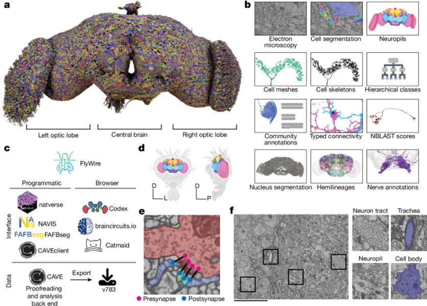
In 2024, the FlyWire consortium reported a neuronal wiring diagram for an adult female Drosophila melanogaster. It reconstructed 139,255 neurons and about 50 million chemical synapses, with annotations for cell types, nerves, hemilineages, and predicted neurotransmitter identities 2. Because the dataset spans both brain hemispheres and includes sensory inputs and motor or endocrine outputs, researchers can trace pathways across larger portions of the animal’s nervous system.
The value is not only descriptive. By following routes from photoreceptors toward descending motor pathways, the researchers generated testable hypotheses about how visual signals could influence behavior 2. A circuit diagram can reveal candidate mechanisms, but it does not prove that every mapped connection is active during a behavior. FlyWire reports that small connections remain underdetected and that synapse prediction performance varies across neuropils and cell types 2. Those qualifications are essential: a connectome is a strong structural constraint, not a complete dynamic simulation.
A mammalian circuit with structure and activity
The MICrONS project brings structural and functional measurements together in mouse visual cortex. Researchers first recorded activity from neurons while mice viewed movies and other visual stimuli. They then processed the same cubic millimeter of tissue with serial electron microscopy and reconstructed the cells and synapses in three dimensions. The resulting resource contains more than 200,000 cells, four kilometers of axons, and 523 million synapses, with a total data volume reported at about 1.6 petabytes 3.
This pairing changes the scientific question. Instead of asking only where a neuron connects, researchers can ask whether neurons with related response properties preferentially connect, how inhibitory cells select their targets, and which anatomical features predict activity. The MICrONS publications report selective inhibitory organization and a wiring preference among neurons with similar response properties 4. The open MICrONS Explorer lets researchers inspect structural reconstructions alongside functional data from the same biological system 5.

Human tissue shows the data challenge
Whole-brain, synapse-level mapping is not yet available for a human brain. A major 2021 Google Research project reconstructed roughly one cubic millimeter of human cortical tissue at nanoscale resolution. The dataset was rendered at about 1.4 petabytes and contained approximately 57,000 cells and 150 million synapses 6. The sample was a small piece of tissue removed during surgery, not a complete brain, and its biological context differs from a living, intact organ.
The scale provides a useful reality check. If a tiny human sample already requires petascale storage and demanding computation, a complete human connectome would require much larger imaging, data-management, and validation systems. The obstacle is not only storage. Researchers must preserve tissue, maintain image continuity, reconstruct branching processes, detect contacts, classify cells, and relate structure to activity across many spatial and temporal scales.
From connectomes to cognitive simulations
A structural map can serve as the substrate for a computational model. Researchers may assign membrane properties, synaptic strengths, delays, neuromodulatory states, and plasticity rules to the reconstructed network. They can then perturb a circuit, replay a stimulus, or compare alternative explanations for a behavior. These experiments are useful because they separate hypotheses that may look similar in a living animal.
A second approach uses data-driven foundation models. In 2025, Stanford researchers described an AI model trained on more than 900 minutes of visual-cortex recordings from eight mice. The model predicted responses to new videos and static images and, for one mouse, inferred anatomical features, cell types, and connections that were checked against high-resolution microscopy 7. The model is not a digital copy of the entire mouse brain. It is a predictive model of a specific visual system, trained on selected data and tested within defined conditions.
The phrase digital twin is useful only when its scope is stated clearly. In neuroscience, current digital-twin work often combines MRI, functional imaging, diffusion measurements, clinical records, and mathematical network models. Such systems can simulate regional activity or explore treatment scenarios, but they simplify the underlying biology 8. A neuron-level connectome and a cognitive digital twin are related but different objects: one emphasizes physical wiring; the other aims to predict behavior or brain dynamics.
What the maps can and cannot tell us
Neuron-level cartography can expose motifs that are hard to detect with coarse imaging. It can reveal rare strong inputs, recurring inhibitory patterns, cell-type-specific routes, and long-range pathways. It also supports reproducible computational experiments: a community can query the same public dataset, rerun an analysis, and compare a model’s prediction with observed activity.
Yet structure does not specify every state of a brain. Synaptic efficacy changes with neuromodulators, learning, sleep, injury, and recent activity. Electrical properties, gene expression, glial interactions, hormones, and body feedback all shape computation. A static map is therefore closer to a detailed coordinate system than to a finished theory of cognition.
There are also ethical questions. Human neural datasets may contain sensitive information about health, identity, or vulnerability, even when they do not record private thoughts in a simple readable form. Governance should cover consent, access, re-identification risk, model ownership, and the use of predictions in medicine or employment. A model that predicts a response is not automatically entitled to make a decision about a person.
Closing Thoughts
The most important shift is methodological. Neuroscience can now connect three kinds of evidence that were often studied apart: anatomy, activity, and computation. That connection does not remove uncertainty, but it makes uncertainty more measurable. It gives researchers a way to state which part of a circuit is observed, which part is inferred, and which part remains a hypothesis.
The strongest future will not come from treating a map as a complete brain. It will come from combining accurate maps with live physiology, behavior, molecular data, and models that can be challenged by new experiments.
What is the true implication of this?
The deeper implication is that complex biological computation is becoming experimentally addressable at the level of its physical components. Researchers can move from broad statements about “brain regions” toward explicit questions about particular cell classes, synapses, pathways, and transformations of information.
That does not mean that consciousness or intelligence has been reduced to a wiring diagram. It means that more of the intermediate mechanisms between cells and behavior can be measured, reconstructed, and tested instead of described only by analogy.
Why does this make a difference?
It makes a difference because a public, queryable circuit map can turn neuroscience into a more cumulative science. A lab can test a prediction against an existing reconstruction rather than starting from a new animal each time. A simulation can expose a weak assumption before it becomes an expensive experiment. A disease model can be compared with a healthy reference at the level of cell types and connections.
The practical promise is conditional: maps must be accurate enough, models must be validated against real biological outcomes, and human data must be governed responsibly. Under those conditions, neural digitalization can become a disciplined bridge between biological observation and computational experimentation.
References
Comments